For severe valgus knee deformity, advantages of the anterolateral procedure are: (1) the lateral release, most usually necessary in valgus knees, is part of the approach. In the alternative case of medial arthrotomy, the vascular supply of the extensor mechanism is seriously impaired; (2) the lateral approach facilitates the release of the lateral contracted elements, offering better surgical view; and (3) the possibility to medicalize the tubercle, if required, improving this way the patellar tracking.
Keblish’s lateral approach comprises of 6 steps:
Step Ⅰ. IT Band Release or Lengthening
Step Ⅱ. Lateral Arthrotomy Coronal Plane Zplasty
Step Ⅲ. Patella DislocationJoint Exposure
Step Ⅳ. Tibial Sleeve Release Osteoperiosteal
Step Ⅴ. Femoral Sleeve Release Osteoperiosteal
Step Ⅵ. Soft Tissue(Prosthetic Joint) Closure
Coronal Plane Zplasty:
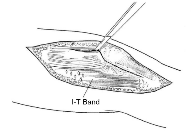
Fig.1
The course of the lateral parapatellar incision begins 24cm lateral to the patella and
extends distally into the midportion of Gerdy’s tubercle (Fig.4.29), preserving the fibrous
layer, which joins with the patellar tendon sheath anteriorly. Proximally, the incision
extends into the central quadriceps tendon.
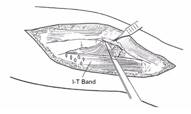
Fig.2
The superficial layer of the retinacular is separated from the deep layer with a coronal
plane Zplasty, from superficial lateral to deep medial. The VL tendon is substantially
thick (610mm) and allows for a horizontal (coronal) plane expansion release. The VL
tendon incision begins near the musculotendonous junction and ends at the mid�
coronal plane of the patella insertion.
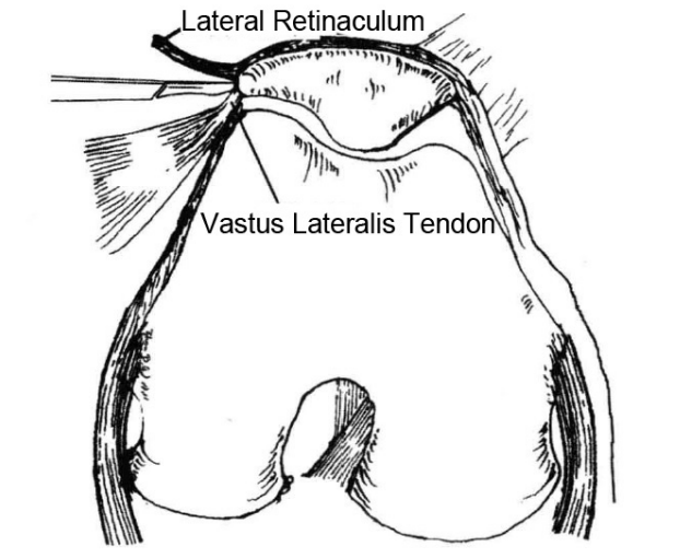
Fig.3
The midportion (lateral retinaculum) separates naturally from the deep capsule and fat pad.
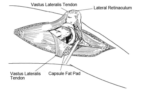
Fig.4
The capsule is incised from the patella rim. The fat pad incision continues
obliquely to the intermeniscal ligament, retaining about 50% of the fat pad with the
patella tendon and 50% with the lateral sleeve, which includes the lateral
meniscus rim for increased soft tissue stability.
Soft Tissue (Prosthetic Joint) Closure:
Soft tissue closure is completed with the knee flexed. The expanded lateral soft tissue sleeve (coronal Zplasty) is positioned to the medial sleeve. Towel clips or stay sutures are utilized, and the knee is extended and flexed through the maximum range. Distal to proximal closure is recommended.
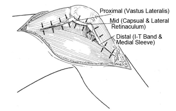
Fig.5
Distally, anatomic reattachment of the IT band and posterolateral sleeve to
Gerdy’s tubercle and the medial sleeve stabilizes the posterolateral corner. In the
midsegment, the capsule is sutured to the lateral border of the retinaculum.
Proximally, the vastus lateralis tendon is reattached in the expanded (coronal
plane Zplasty) position with the knee in maximal flexion. The knee is ranged to a
full flexion position (140 to 150 degrees), and soft tissue compliance and integrity
of the joint seal are checked. Appropriate adjustments or reinforcing sutures can
be implemented at this time. The preoperative noncompliantdeforming lateral
soft tissue structures are now more compliant and allow for coverage of the
prosthetic joint.
 Mar. 17, 2020


